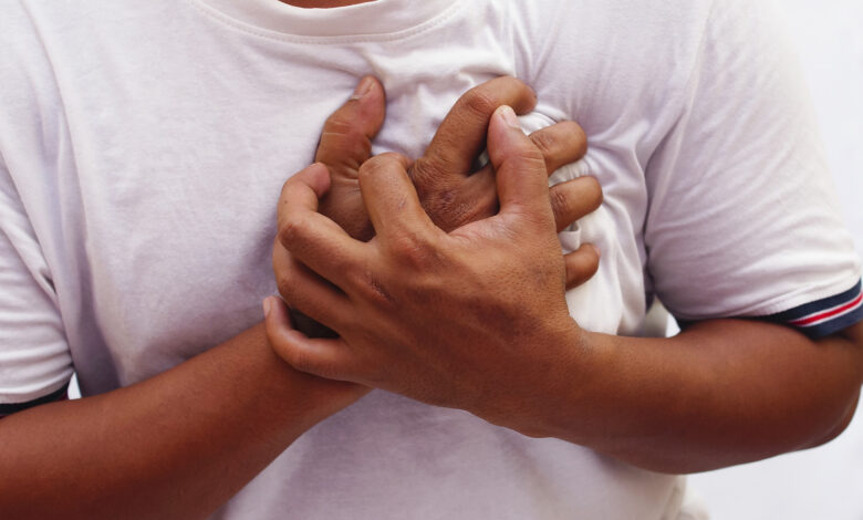
Case three is a 66 year old male smoker who presents to the ER with 24 hours of chest pain. He has not had this pain before and describes it as a burning and heaviness in the breast bone area. The pain radiates up to his neck, left jaw, shoulder and arm, and is accompanied by nausea, perspiration, and difficulty breathing. It worsened after an argument with his son.
On exam, he appeared distressed and uncomfortable. His skin was pale and clammy. Heart rate was 90, BP 110/70. His lungs were clear. He had a slight heart murmur heard along the left border of his breast bone. Abdomen and extremities are normal.
An electrocardiogram was done immediately and showed changes that indicated there was acute injury to the front (anterior) wall of his heart. An EKG normally has 12 “leads” which look at the heart from 12 different views, or positions. “Leads” V1 through V6 capture what’s going on in the front (anterior) wall of the heart. On this man’s EKG, leads V1 through V5 had changes indicating there was acute injury to his anterior heart wall. The term used is ST segment elevation.
He was immediately taken to the cardiac catheterization lab for coronary angiography. “Dye” was injected into his right and left coronary arteries to see if and where there was a blockage of coronary blood flow. It showed a clotted area obstructing flow in the left anterior anterior descending artery (LAD), a major branch of the left coronary artery. Evidence of heart injury beyond the blockage was seen. He underwent a balloon angioplasty (re-opening of the blockage by stretching it) and stent placement to keep the blockage from closing off again.
The procedure was a success, the patient’s chest pain went away, he could breathe easier, and he felt better. He was stable after the procedure, and the next day was released to be seen in the office in a week.
What’s wrong with this patient? He has a blockage of the left anterior descending coronary artery and an acute anterior wall myocardial infarction: a heart attack.
Emergency angiography and stenting are done to minimize the amount of damage done to the heart muscle by re-establishing blood flow to the damaged area. Getting oxygen to the heart muscle is very important for preserving damaged myocardium (heart muscle). Preserving myocardium is important for maintaining physical activity and preventing heart failure.
This is a fairly straightforward case and is a good example of why patients no longer spend three weeks in the hospital on bed rest after a heart attack. Emergency angiography, angioplasty, and stenting save lives, preserve myocardium, and lessen arrhythmias. And it keeps cardiologists very busy.
Reference: Ribeiro WN, Yamada AT, Benvenuti LA. Case 4/2014-A 66-year-old-man with Acute Myocardial Infarction [and death in asystole after primary coronary angioplasty] Arq Bras Cardiol 2014 Sep;103(3):e31.doi:10.5935/abc.20140129