The Coronary Arteries are the arterial vessels that supply oxygen-rich blood to the myocardium, the heart muscle. Oxygen must be continuously supplied to the heart muscle or the muscle will infarct, or die. That is the definition of a heart attack: due to a partial or complete blockage of a coronary artery, the heart muscle’s demand for oxygen exceeds the coronary’s ability to supply oxygen, so the heart muscle dies. It called an Ischemic Myocardial Infarction.
There are two coronary arteries, both of which originate in the base of the aorta just above the aortic valve. A brief review of the anatomy of the heart tells us the heart has 4 chambers— the Right Atrium (RA) and Right Ventricle (RV), and the Left Atrium (LA) and Left Ventricle (LV). The RA and RV are connected by the Tricuspid Valve. The LA and LV are connected by the Mitral Valve. Blood exits the RV through the Pulmonic Valve and flows into the lungs to get oxygenated. Blood from the lungs flows to the LA through the Mitral Valve into the LV then exits through the Aortic Valve to the entire body. Oxygenated blood leaving the LV through the Aortic Valve also flows into the Right Coronary (RCA) and Left Coronary (LCA) arteries, giving life and function to the myocardium. The atria and ventricles are separated by a membrane called the septum.
The LCA begins as a single artery (the Left Main (LM)artery) but quickly branches into the Left Anterior Descending Artery (LAD) and the Left Circumflex (LCx) Artery.
The LAD supplies the front, the bottom left, and two-thirds of the septum between the ventricles; a large area. It is nicknamed “the widow maker” because when it is blocked, a large area of myocardium is affected.
The LCx artery wraps around the left side of the heart supplying blood to the LA.
The LAD branches into the Diagonal and Septal branches.
The LCx branches into the Obtuse Marginal (OM) which supplies the side and back walls of the LV.
The RCA is usually a single artery, and it supplies the RA, the RV, and the SinoAtrial (SA) node where all heart beats originate.
The RCA has 4 branches-the Conus, the Acute Marginal, the Posterior Descending (PDA), and the AV Nodal branches. The PDA supplies the bottom, back, and septum on the Right side.
EKG changes tell doctors where the blockage has occurred and if it is totally or partially blocked. An EKG has 12 leads or terminals that connect to the body. They are I, II, III, AVR, AVL, AVF, and V1-6.
When an artery is totally blocked, the EKG shows ST segment elevation in the area supplied by that artery.
When artery is partially blocked, the EKG shows ST segment depression and T wave inversion
Below, we will see what EKG leads are affected by a blockage in each artery:
LAD blockage—V1-4 show ST elevation, II, III, AVF show ST depression.
RCA blockage—ST elevation in II, III, AVF and ST depression in lateral and back leads I, AVL
LCx blockage—ST elevation in I, AVL, V5-6. For a posterior blockage ST depression in V1-3
LM blockage—ST depression in V2-6, II, III, AVF
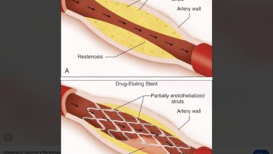
I know this is Greek to most readers, and I apologize for that. But this is a good overview of Coronary Artery anatomy and the EKG changes that occur when specific arteries are blocked. The main takeaway message is there are two coronaries with multiple branches. Disease in the LM, LAD, LCx, or at the beginning of the RCA, has the most serious effect on the patient. Early, aggressive Angioplasty and stenting of any blockage is a life-saving measure.
Reference: www.google.com/view-article/coronary-artery-anatomy
Smithuis R, Willems T. Coronary anatomy and anomlies. Rad Asst 2008 Oct 14.
www.google.com/view-article/EKG-Changes-affect-leads.